
Figure 3
- ID
- ZDB-FIG-230321-13
- Publication
- Xiao et al., 2023 - Dopey2 and Pcdh7 orchestrate the development of embryonic neural stem cells/ progenitors in zebrafish
- Other Figures
- All Figure Page
- Back to All Figure Page
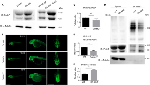
Dopey2 modulates the ubiquitination of Pcdh7b protein (A) Left panel illustrates immunoblotting detected Pcdh7 protein levels in 48 hpf WT embryos pre-injected with Ctl MO or DO MO; right panel illustrates immunoblotting detected Pcdh7 protein levels in 48 hpf WT or DO MUT embryos. Immunoblotting of α-tubulin was used as loading control. (B) Whole-mount immunofluorescence staining detected Pcdh7 protein in Ctl MO or DO MO injected WT embryos and DO MO embryos at 48 hpf. Scale bar = 500 μm. (C) Real-time fluorescence quantitative PCR analysis detected relative expression level of pcdh7b in WT or DO MUT embryos. (mean ± s.e.m, n = 3, Student’s t test: NS = not significant). (D) Co-immunoprecipitation (co-IP) analysis detected the ubiquitination of Pcdh7 in 48 hpf WT or DO MUT embryos. IP: immunoprecipitation, IB: immunoblot, Ub: anti ubiquitin antibody. (E) Quantification of immunoprecipitated Pcdh7 (DO MUT embryos) indicated a reduction in ubiquitination (mean ± s.e.m, n = 3, Student’s t test: ∗p < 0.05). Images were quantified by ImageJ software. (F) Quantification of immunoprecipitated Pcdh7 (DO MUT embryos) indicated Pcdh7 had an increase at protein level in lysates (mean ± s.e.m, n = 3, Student’s t test: ∗p < 0.05). Images were quantified by ImageJ software. Ctl MO: control MO, DO MO: dopey2 MO, WT: wild type, DO MUT: dopey2mutant. |