
Fig. 3
- ID
- ZDB-FIG-230226-3
- Publication
- Slováková et al., 2022 - Tension-dependent stabilization of E-cadherin limits cell-cell contact expansion in zebrafish germ-layer progenitor cells
- Other Figures
- All Figure Page
- Back to All Figure Page
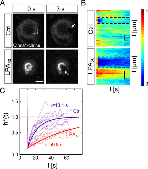
Cortical tension reduces Ctnna1 turnover at the contact of cell doublets. (A) FRAP analysis of Ctnna1 turnover at the contact of progenitor cell doublets. Fluorescence images of Ctnna1 localization within the contact plane of control doublets (Upper) and doublets exposed to 50 nM LPA (Lower) in the last prebleach (Left) and first postbleach frames (Right). Boxed regions (Left) and arrows (Right) outline bleached regions. (Scale bar: 10 μm.) (B) Normalized intensity kymographs of Ctnna1 recovery after photobleaching at the cell–cell contact edge of control doublets and doublets exposed to 50 nM LPA. (Scale bars: horizontal, 6 s; vertical, 10 μm.) (C) Quantification of Ctnna1 fluorescence intensity within the bleached regions at the contact edge of control doublets (purple) and doublets exposed to 50 nM LPA (red) as a function of time after photobleaching. τ denotes the recovery characteristic timescale. Thin lines denote individual cases, and thick lines are averages. (Ctrl) N = 3 and n = 12; (LPA50) N = 2 and n = 7. Materials and Methods has details. If not stated otherwise, N corresponds to the number of experiments, and n corresponds to the number of cell doublets. |