|
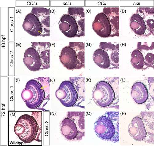
Loss of Crk adaptor proteins leads to retinal lamination defects. Transverse sections of zebrafish embryos stained with Hematoxylin and Eosin through the center of the forebrain, including the retina, of CCLL (A, E, I), ccLL (B, F, J, N), CCll (C G, K, O), and ccll (D, H, L, P) at 48 hpf (A–H) and 72 hpf (I–P). For wild-type comparison, retina section of a 72 hpf Sofa1 transgenic embryo (M),30 the genetic background of the mutant lines. Embryos were classified by retinal lamination phenotype based on % inner plexiform layer (IPL) per total outer curve (48 hpf) and % outer plexiform layer (OPL) per total outer curve (72 hpf) measurements (see Figure 7 for quantification, yellow arrows in A and I indicate IPL and OPL in 48 hpf and 72 hpf, respectively), and sample images of each class are shown: 48 hpf class 1 (A–D), 48 hpf class 2 (E–H), 72 hpf class 1 (I–L), and 72 hpf class 2 (N–P)
|