
FIGURE 2
- ID
- ZDB-FIG-221227-8
- Publication
- Palma et al., 2022 - Ontogenesis of the asymmetric parapineal organ in the zebrafish epithalamus
- Other Figures
- All Figure Page
- Back to All Figure Page
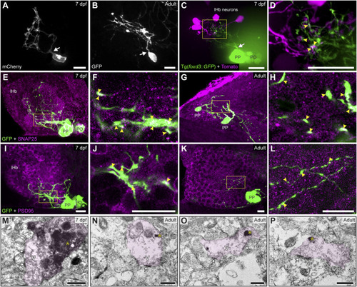
Contacts and synapses between parapineal cells and the left habenular neuropil in the larva and adult zebrafish. (A,B) Dorsal views of confocal z-stack maximum projections showing single parapineal cells expressing mCherry in a living larva at 7 dpf (A) and GFP in a section of the 1.5–2 years old adult brain (B) after immunostaining against this fluorescent protein. At both stages, parapineal cells show a characteristic pear-shape cell body with a single projection emerging from it (white arrows) and then branching profusely. (C,D) Dorsal views of confocal z-stack maximum projections showing 4 habenular neurons expressing the fluorescent protein Tomato (magenta) in a living 7 dpf Tg(foxd3::GFP) larva expressing GFP in the pineal complex (green). Projections emerge from the parapineal body as a bundle (white arrow), then branch in the left habenula to intermingle with the dendritic arbour of the labelled habenular neurons. The image in (D) corresponds to a z-plane of the yellow rectangle depicted in (C), showing the punctuated zones where the fluorescent signals corresponding to parapineal projections (green) closely associate with the fluorescence signals corresponding to habenular dendrites (magenta). (E–L) Dorsal views of immunofluorescence against presynaptic (SNAP25) and postsynaptic (PSD95) markers in the epithalamus of larval (7 dpf) and adult (1.5–2 years old) zebrafish. Images correspond either confocal z-stack maximum projections (E,G,I,K) or to single z-planes (F,H,J,L; corresponding to the yellow rectangles depicted in E,G,I,K, respectively) of larval (E,F,I,J) and adult (G,H,K,L) animals. Yellow arrowheads point to zones where the fluorescent signal from to parapineal projections (green) closely associate with the immunofluorescence signal of the synaptic protein (magenta). (M–P) TEM images showing the synaptic relation between parapinal cells and the habenular neuropil at larval (7 dpf) (M) and adult (1.5–2 years old) (N–P) stages. GFP-immunopositive terminals of parapineal projections showing black DAB precipitates (yellow asterisks) are shaded in pink. These terminals show synaptic vesicles (v) and synaptic densities (white arrows) both in larvae and adults. Abbreviations: lHb (left habenula), PO (pineal organ at the level of the stalk), PP (parapineal). Scales bars, 10 µm (A–L), 500 nm (M–P). |