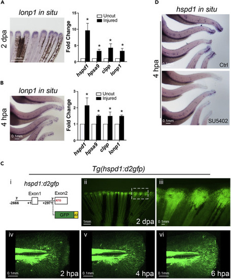
Figure 1. The UPRmt is activated in adult and larval injured fins
(A and B) Representative in situ hybridization images (left panels) show that lonp1 (a UPRmt gene) is up-regulated during fin regeneration in adult (A, partial fin view, posterior toward top) and larval (B, partial body view, posterior toward the right) fish at 2 dpa or 4 hpa, respectively. qRT-PCR assays (right panels) show that the expression of all four UPRmt signature genes are enhanced in adult and larval injured fin tissue.
(C-i) Diagram indicates the hspd1 genomic fragment (2.6 kb upstream of the transcription start (+1) to 3 kb downstream until the translation start) employed for driving the expression of the destabilized GFP. (C-ii and C-iii) Adult fin of the UPRmt reporter fish, Tg(hspd1:d2gfp), exhibits induced fluorescence at the injury zone during regeneration. Higher magnification image (iii; white rectangle in ii) indicates that the fluorescent signal marks the blastema of the injured fin. (C-iv to C-vi) A time series of images shows the dynamics of the UPRmt reporter activation in larval fin upon injury. Note fluorescent migrating mesenchymal-like cells.
(D) Representative images of hspd1 in situ hybridization assays show that hspd1 expression is diminished in SU5402 treated injured larvae at 4 hpa. (Error bars indicate standard deviation. Student’s t test was performed to determine statistical significance. ∗p < 0.05). Scale bars are as indicated.