
Fig. 6
- ID
- ZDB-FIG-221018-215
- Publication
- Yong et al., 2021 - SNX27-FERM-SNX1 complex structure rationalizes divergent trafficking pathways by SNX17 and SNX27
- Other Figures
- All Figure Page
- Back to All Figure Page
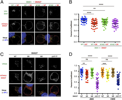
Interaction with SNX1 and VPS26 mediates the endosomal recruitment of SNX27 in Jurkat T cells. Jurkat T cells were transfected with plasmids expressing YFP-SNX1 and mCherry-SNX27 (A and B) or with plasmids expressing mCherry-SNX27 (C and D) as indicated and conjugated with NALM6 B cells. (A) Representative images showing localization of SNX1 (green) and SNX27 (red) in T cells conjugated with staphylococcal enterotoxin E (SEE)-pulsed B cells (APC; blue). (B) Colocalization analysis between SNX1 and SNX27 within T cells conjugated with SEE-pulsed B cells. (C) Representative images showing localization of SNX27 (red) and endogenous VPS35 (green) in T cells conjugated with SEE-pulsed B cells (APC; blue). (D) Colocalization analysis between SNX27 and VPS35 within T cells conjugated with NALM6 B cells preloaded with or without SEE superantigen. (A and C) (Scale bar: 5 μm.) (B and D) Each dot represents a Pearson’s correlation coefficient from a single conjugated T cell, and error bars indicate SD. Data shown are representative (A and C) and combined (B and D) from four (A and B) and three (C and D) independent experiments. Statistical significance was determined using one-way ANOVA with Tukey’s multiple comparisons test. **P < 0.01, ***P < 0.001, ****P < 0.0001, ns, not significant. |