
Fig. 1
- ID
- ZDB-FIG-220927-86
- Publication
- Naylor et al., 2022 - A novel nanoluciferase transgenic reporter measures proteinuria in zebrafish
- Other Figures
- All Figure Page
- Back to All Figure Page
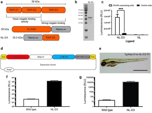
Figure 1. NL-D3 is uptaken by the megalin endocytosis pathway. (a) The top schematic shows binding affinity to megalin of the 3-dimensional domains of the receptor-associated protein (RAP). The bottom schematics show the size and orientation of full-length RAP bound to the N-terminus of Nano-Luc and the RAP D3 domain bound to the C-terminus of Nano-Luc (NL-D3, which was used in this work). (b) Sodium dodecylsulfate-polyacrylamide gel electrophoresis gel showing the recombinantly expressed and purified NL-D3 protein alongside molecular weight markers. (c) Graph showing the internalization of NL-D3 or untagged NL into MmR4 mini-megalin expressing cells and control nonmegalin expressing cells. ∗∗∗P ≤ 0.001. (d) Schematic for the γ-crystallin:mcherry/fabp10a:NL-D3 Tol2 vector used to generate transgenic zebrafish. (e) Panel showing the lateral view of a 5-day post-fertilization (dpf) NL-D3 transgenic zebrafish embryo under excitation for red fluorescence to highlight the mCh expression in the lens. Bar = 1 mm. (f) Histogram showing relative luminescence units (RLU) in whole embryo lysates of wild-type and NL-D3 5 dpf embryos. (g) Logarithmic histogram showing the RLU of 1 μl blood from adult wild-type and NL-D3 zebrafish. |