
Fig. 3
- ID
- ZDB-FIG-220912-114
- Publication
- Wang et al., 2022 - Tankyrase Inhibition Attenuates Cardiac Dilatation and Dysfunction in Ischemic Heart Failure
- Other Figures
- All Figure Page
- Back to All Figure Page
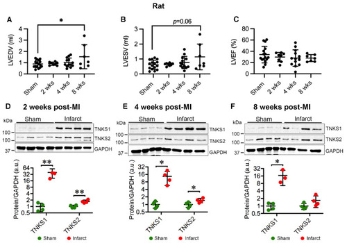
Ischemic injury boosts TNKS1 and TNKS2 in the infarct area in rat hearts. (A,B) Echocardiography depicts increased left ventricular volume at 8 weeks post-MI compared to sham controls as determined by left ventricular end diastolic volume (LVEDV) (A) and left ventricular end systolic volume (LVESV) (B). (C) Left ventricular ejection fraction (LVEF). (D–F) Representative immunoblots of TNKS1 and TNKS2 in the infarct area in MI rat hearts and sham-operated controls at 2 (D), 4 (E), and 8 (F) weeks post-MI. The graphs represent quantifications of three replicate blots for the indicated proteins normalized to GAPDH. The results are presented as fold change compared to controls. The blurry band was excluded from the quantification. (A–C) Sham, n = 16; 2 weeks, n = 8; 4 weeks, n = 13; 8 weeks, n = 7. (D), Sham, n = 4; MI, n = 4. (E) Sham, n = 4; MI, n = 4. (F) Sham, n = 4; MI TNKS1, n = 3; TNKS2, n = 4. Data are presented as mean ± SD. One-way ANOVA with Tukey adjustment for multiple comparisons (A–C); two-sample t-test (D–F); * p < 0.05, ** p < 0.01. |