|
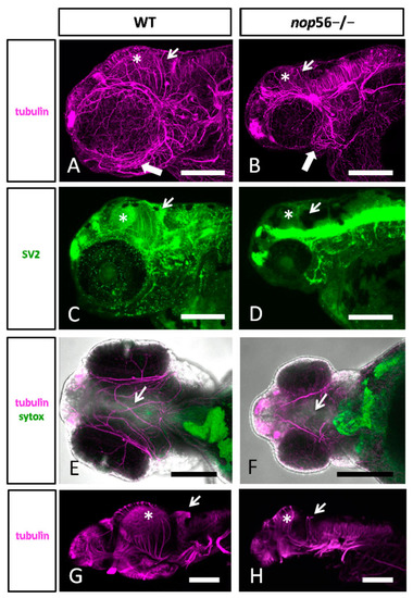
(A,B) Lateral views of WT (A) and nop56−/− (B) 3.5 dpf larvae immunostained against α-tubulin (magenta) showing abnormalities in the jaw (thick arrow) and brain, specially in the midbrain (asterisk) and cerebellum (thin arrow). (C,D) Lateral views of WT (C) and nop56−/− (D) fish immunostained against SV2 (green) showing different distribution in the brain, especially in the midbrain (asterisk) and cerebellum (arrow). (E,F) Ventral views of WT (E) and nop56−/− (F) showing lack of midline fibers and bundles in the homozygous fish (arrow in (F)), compared to WT (arrow in (E)). (G–F) Lateral views of WT (G) and nop56−/− (H) brain showing smaller midbrain in the homozygous fish (asterisk in (H)) and absence of labelling in cerebellum (arrow in (H)), compared to WT (asterisk and arrows in (G)). All images are projections from confocal z-stacks. Scale bars: 125 μm.
|