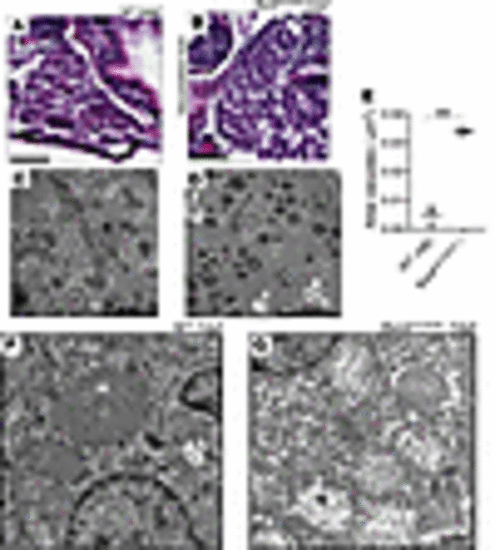
Biochemical analysis, histopathology, and ultrastructural investigation of homozygous fbxl4sa12470 zebrafish liver. (A) Liver histology in AB (WT) zebrafish larvae at 6 dpf. Scale bar: 25 μm. (B) Vacuolated liver in homozygous fbxl4sa12470 zebrafish larvae at 6 dpf. 100% of larvae (n = 25) showed vacuolated liver, while no WT larvae (n = 12) showed the disease phenotype. Scale bar: 25 μm. (C) Ultrastructure of hepatocytes in 7 WT dpf zebrafish larvae showing normal ultrastructure. Scale bar: 10 μm. (D) Ultrastructure of hepatocytes in mutant 7 dpf zebrafish larvae showing increased rate of lipid droplets (L) and autophagic vacuoles (white arrows). Scale bar: 10 μm. (E) Total area of vacuoles occupying liver areas estimated in μm2 in mutant versus WT larvae at 7 dpf. Total area of liver analyzed: 1,978 μm2 (WT) and 2,741 μm2 (fbxl4sa12470). **P < 0.01. Significance was determined using unpaired t test. n = 3 each fish line. (F) Mitochondrial ultrastructure (M) in hepatocytes of 7 dpf WT zebrafish larvae. (G) Loss of normal matrix electron density and mitochondrial cristae damage (M) was observed in hepatocytes of 7 dpf fbxl4sa12470–/– larvae. Scale bar: 1 μm (F and G).
|
