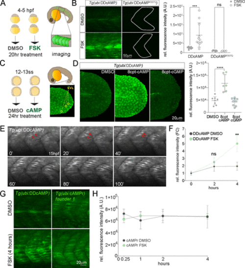
Figure 3. (A) Diagram of DMSO and 20 μM FSK treatment on Tg(ubi:DDcAMP) and Tg(ubi:DDcAMPR307Q) embryos beginning at 4–5 hpf for 20 h. Anterior somites from treated embryos were imaged at 24 hpf, and GFP intensity was measured in somites 7–11. (B) Images of somites from DMSO and FSK-treated Tg(ubi:DDcAMP) and Tg(ubi:DDcAMPR307Q) embryos at 24 hpf. The graph represents the mean fluorescence intensity of five somites per embryo, and each point corresponds to one embryo. Error bars indicate SD: ***p < 0.001 by two-way ANOVA (Šídák’s multiple comparisons), n = 6–11 animals for each condition. ns = not significant. AU = arbitrary unit. (C) Diagram of DMSO and 8-cpt-cAMP treatment of Tg(ubi:DDcAMP) starting at 12–13 somite stage (12–13ss) and imaged after 24 h of incubation. The tip of the tail was imaged for GFP intensity and the EVLs were quantified. (D) Tg(ubi:DDcAMP) embryos from the same clutch were treated with DMSO, 100 μM 8-cpt-cAMP, or 100 μM 8-cpt-cGMP; signal from 20 EVL cells was averaged per animal. Each dot in the graph represents one animal. Error bars indicate SD; ****p < 0.0001 one-way ANOVA (with Bonferroni’s multiple comparisons), n = 6–9 animal per condition. AU = arbitrary unit. (E) Frames from a confocal time lapse image with Airyscan 2 processing of Tg(ubi:DDcAMP) embryo starting at 15 hpf (t = 0 min). One frame every 20 min is shown as representation of the time lapse. GFP-expressing muscle cells are indicated with red arrows. (F) Graph represents the fold change of mean fluorescence intensity measured in five somites per embryo at time 0 and after 2 and 4 h of treatment. Error bars indicate SEM; **p < 0.01 two-way ANOVA (with Šídák’s multiple comparisons), n = 6–10 animals for each condition. ns = not significant, FC = fold change. (G) Confocal images of somites from Tg(ubi:DDcAMP) and Tg(ubi:cAMPr) embryos after 4 h of FSK and DMSO treatment show GFP expression in muscle cells but different response to FSK treatment. (H) Graph represents the mean fluorescence intensity measured in five somites per embryo at the time points indicated. Error bars indicate SEM; there is no significance among the groups; n = 4–5 animals for each condition.
|
