
Fig. 7
- ID
- ZDB-FIG-220819-23
- Publication
- Willekers et al., 2022 - The centriolar satellite protein Cfap53 facilitates formation of the zygotic microtubule organizing center in the zebrafish embryo
- Other Figures
- All Figure Page
- Back to All Figure Page
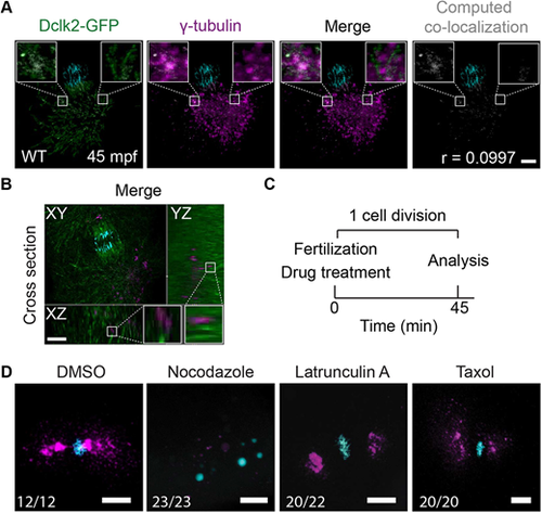
Microtubule-dependent γ-tubulin localization. (A,B) Maximal projections of confocal stacks from a Tg(Xla.Eef1a1:Dclk2a-GFP) embryo immunolabeled for DAPI (in cyan), Dclk2a-GFP (in green) and γ-tubulin (in magenta) at 45 mpf fixed during telophase. Colocalization of γ-tubulin and Dclk2a-GFP is shown in white in the merge. Computation of colocalization gives an average Pearson's coefficient of 0.1, indicating that there is very little colocalization of γ-tubulin and Dclk2a-GFP (A, n=5). Scale bar: 100 μm. (B) Optical cross-section through 45 mpf embryo immunostained for γ-tubulin and Dclk2a-GFP at the region of one of the centrosomes, which shows that γ-tubulin is localized directly adjacent to the microtubule bundles (B, n=5). Scale bar: 100 μm. White squares outline the areas shown in more detail. (C) Schematic overview of the drug treatment experiment (C), for which the results are shown in D. (D) Maximal projections of confocal stacks of embryos at 45 mpf immunostained with γ-tubulin (in magenta) and DAPI (in cyan) fixed during telophase. Nocodazole treatment (n=23) prevents γ-tubulin accumulation at the MTOC. Lantrunculin A (n=20) and taxol (n=20) treatments have no effect on γ-tubulin localization to the MTOC. Scale bars: 20 μm. |