
FIGURE
Fig. 5
- ID
- ZDB-FIG-220819-15
- Publication
- Wu et al., 2022 - dock8 deficiency attenuates microglia colonization in early zebrafish larvae
- Other Figures
- All Figure Page
- Back to All Figure Page
Fig. 5
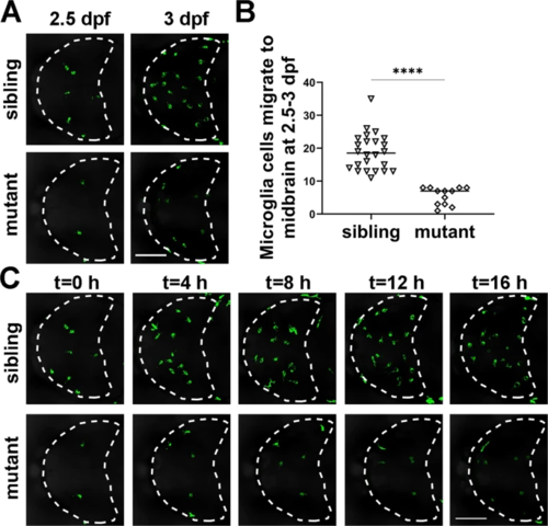
A Representative images of GFP+ microglia in dock8-15,+5bp; Tg(coro1a:GFP) embryos from 2.5 dpf–3 dpf for 10–16 h. B Quantification of GFP+ microglia migrating into the midbrain from 2.5 dpf to 3 dpf in dock8-15,+5bp; Tg(coro1a:GFP) embryos. C Time-lapse confocal imaging of microglia colonization in dock8-15,+5bp; Tg(coro1a:GFP) embryos from 2.5 dpf–3 dpf. Each dot represents one larva. White dotted lines indicate the midbrain. Scale bar = 100 µm. See also Video S2. Data were analyzed by unpaired Student’s t-tests. ****P ≤ 0.0001. |
Expression Data
| Gene: | |
|---|---|
| Fish: | |
| Anatomical Term: | |
| Stage Range: | Pec-fin to Protruding-mouth |
Expression Detail
Antibody Labeling
Phenotype Data
| Fish: | |
|---|---|
| Observed In: | |
| Stage Range: | Pec-fin to Protruding-mouth |
Phenotype Detail
Acknowledgments
This image is the copyrighted work of the attributed author or publisher, and
ZFIN has permission only to display this image to its users.
Additional permissions should be obtained from the applicable author or publisher of the image.
Full text @ Cell Death Discov