
FIGURE
Figure 2
- ID
- ZDB-FIG-220801-170
- Publication
- Somkhit et al., 2022 - Microglia Remodelling and Neuroinflammation Parallel Neuronal Hyperactivation Following Acute Organophosphate Poisoning
- Other Figures
- All Figure Page
- Back to All Figure Page
Figure 2
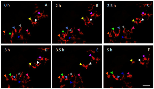
Dynamics of microglia remodelling in DFP-exposed embryos. Selected snapshot views of live imaging of microglia showing the dynamics of microglia morphological changes in a living 5 dpf Tg[mpeg1:mCherryF] larva, before (0 h) and at different time points of a 6 h exposure to 15 µM DFP. Coloured arrowheads indicate individual microglial cells. Scale bar represents 20 µm. |
Expression Data
Expression Detail
Antibody Labeling
Phenotype Data
Phenotype Detail
Acknowledgments
This image is the copyrighted work of the attributed author or publisher, and
ZFIN has permission only to display this image to its users.
Additional permissions should be obtained from the applicable author or publisher of the image.
Full text @ Int. J. Mol. Sci.