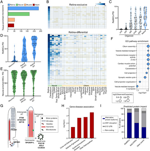
Characterization and identification of the human RetMIC program. (A) Number of tissue-enriched microexons by tissue type in humans. Only the four tissues with the highest number of tissue-enriched microexons are depicted. (B) Heatmap showing RetMIC inclusion in different human tissues. RetMICs are divided into retina-exclusive (inclusion only in retina samples) and retina-enriched (biased inclusion in retina compared to neural samples). Each row corresponds to a different microexon. Inclusion levels were obtained from VastDB (23). (C) Inclusion levels (PSIs) of RetMICs in cone-rich human organoids (SRP056957) and whole retina. Developing organoid time points: day 15 (d15), day 30 (d30), day 85 (d85), day 194 (d194), and day 250 (d250). Data plotted are averaged PSIs of three biological replicates; events with NA values due to insufficient read coverage were omitted. (D and E) Violin plots depicting inclusion levels of mouse RetMICs (D) and neural-enriched microexons (neural MICs) (E) in hippocampal neurons, WT and Aipl1 KO retinae (data from SRP068974). Events with insufficient read coverage were omitted. (F) Top 10 enriched GO terms for human RetMIC-containing genes. P values are false-discovery rate (FDR)-adjusted. (G) Schematic representation of the OS and localization of selected RetMIC genes; IFT, intraflagellar transport. (H) Enrichment of RetMIC-containing genes among loci associated with different retinal diseases. P values from hypergeometric tests. Complete inputs and results are provided in Dataset S3. (I) Predicted protein impact of different exon types as annotated in VastDB.
|
