
Fig. 2.
- ID
- ZDB-FIG-220717-57
- Publication
- Casey et al., 2021 - Shutdown corner, a large deletion mutant isolated from a haploid mutagenesis screen in zebrafish
- Other Figures
- All Figure Page
- Back to All Figure Page
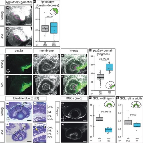
Optic cup patterning is partially altered and retinal defects arise in shutdown corner. a–c) Expression of Tg(cldnb:lyn-EGFP), which labels the nasal (anterior) hemisphere of the optic cup at 24 hpf. 3D-rendered, lateral view of Tg(cldnb:lyn-EGFP);Tg(bactin2:EGFP-CAAX) embryos. a, b) Magenta lines demarcate Tg(cldnb:lyn-EGFP)-positive region. c) Quantification of Tg(cldnb:lyn-EGFP)-positive domain per embryo. d–j) Antibody staining for pax2a, a ventral marker, at 24 hpf. 3D-rendered, lateral views. Pax2a (d, g; green), cell membranes (e, h; grayscale, EGFP-CAAX), merge f, i). Magenta lines f, i) demarcate pax2a-positive region. j) Quantification of the pax2a-positive domain per embryo. k, l) 5 dpf histological sections stained with toluidine blue (imaged at 40x, sections at similar depth based on presence of optic nerve; sibling n = 2, sco n = 3) with zoomed views of the retina (k′, l′). Arrows, optic nerve; bracket, photoreceptor outer segments k′); asterisk, missing photoreceptor outer segments l′); arrowhead, potential cell death l′). m–p) RGCs (zn-5 staining) at 5 dpf. Ventral view, single confocal section from 3D datasets of antibody-stained samples. o, p) Quantification of GCL thickness, presented as the raw width o) and the width normalized to the total width of the retina p). Width measurements were taken at three places in each retina, at a nasal, temporal, and nasal-temporal midpoint; each point represents the average GCL width (raw or normalized) per embryo. ON, optic nerve; ONL, outer nuclear/photoreceptor layer; INL, inner nuclear layer; IPL, inner plexiform layer; GCL, ganglion cell layer; RGC, retinal ganglion cell; A, anterior; D, dorsal; L, lateral; M, medial; ON, optic nerve; P, posterior; V, ventral. Scale bar, 50 µm. |