
Fig. 1
- ID
- ZDB-FIG-220527-18
- Publication
- Xiao et al., 2022 - Identifying drivers of breast cancer metastasis in progressively invasive subpopulations of zebrafish-xenografted MDA-MB-231
- Other Figures
- All Figure Page
- Back to All Figure Page
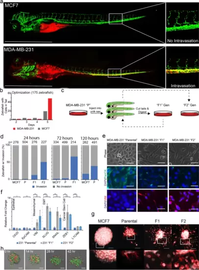
Labeled MDA-MB-231 cells were injected into the yolk sac of 2-days post fertilized zebrafish embryos and monitored for invasion. a Injection of MCF7 and MDA-MB-231 cells into the yolk sac resulted in cell arrest within the caudal plexus. Images of zebrafish were taken at 4x magnification using Olympus IX-71 inverted fluorescence microscope. b Transiently labeled cells arrest within the tail of the zebrafish within 5 days of injection. c Workflow of serial transplantation of the MDA-MB-231 heterogeneous parental population to generate the F1 and F2 subpopulations. d MDA-MB-231 F1, and F2 cells arrest within the tails of zebrafish progressively faster. Three separate experiments evaluating the parental (“P”), F1, and F2 ability to invade over three time points (120, 72, and 24 hours). Xenografted parental, F1, and F2 cells appeared in ≥40% of injected zebrafish in 120, 72, and 24 hours, respectively. e Phase and immunofluorescence images of resulting in vitro cultured MDA-MB-231 parental, F1, and F2 cells. f qRT-PCR amplification of epithelial (CK20, EpCAM), mesenchymal (VIM), EMT TFs (SLUG, SNAIL), and cancer stem cell markers (ZEB1, L1CAM) was performed of the three subpopulations. Relative fold changes were normalized to MDA-MB-231 parental cells. * = p-value < .05; ** = p-value < .001; *** = p-value < .0001 (g) Clusters of MCF7, MDA-MB-231 parental, F1, and F2 cells were embedded within a 3-dimensional Matrigel-based extracellular matrix and allowed to invade over 24 hours. Clusters were stained for nuclei (white) and phalloidin (red). h RFP-labeled F2 and GFP-labeled parental cells were co-clustered and embedded in 3D ECM showing F2 cells invade the ECM progressively with time, but not parental cells. Scale bars = 100 μm |
| Gene: | |
|---|---|
| Fish: | |
| Condition: | |
| Anatomical Term: | |
| Stage: | Day 5 |