
FIGURE
Figure 4
- ID
- ZDB-FIG-220430-125
- Publication
- Džulová et al., 2022 - Incomplete Recovery of Zebrafish Retina Following Cryoinjury
- Other Figures
- All Figure Page
- Back to All Figure Page
Figure 4
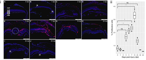
Figure 4. PCNA staining of proliferation: (i) (A) Proliferation is minimal in the control uninjured retina (0 dpi). (B) 1 dpi: proliferation begins to be detected (arrows). (C,D) 2 dpi and 3 dpi: proliferation more evident at the lesion site. (E) 5 dpi: proliferation pronounced, with positive nuclei located predominantly in the ONL at the lesion margin, absent in the lesion site (circled). (F) 7 dpi: proliferation now detected throughout the lesion. (G,H) 10 dpi and 20 dpi: proliferation started to decrease as new cells have started to repopulate the lesion site. (I,J) 30 dpi and 60 dpi: proliferation now only marginally detected (arrows). All sections were counterstained with DAPI. PCNA proliferation marker—red; Nuclei, DAPI—blue. Retinal layers labelled in control panel for reference; RPE—retinal pigment epithelium, ONL—outer nuclear layer, OPL—outer plexiform layer, INL—inner nuclear layer, IPL—inner plexiform layer, GCL—ganglion cell layer. Scale bars represent 200 µm. (ii) Relative percentage of proliferative cells at each time-point (n = 2), * p value < 0.05.
|
Expression Data
Expression Detail
Antibody Labeling
Phenotype Data
Phenotype Detail
Acknowledgments
This image is the copyrighted work of the attributed author or publisher, and
ZFIN has permission only to display this image to its users.
Additional permissions should be obtained from the applicable author or publisher of the image.
Full text @ Cells