
FIGURE
Figure 1
- ID
- ZDB-FIG-220430-113
- Publication
- Džulová et al., 2022 - Incomplete Recovery of Zebrafish Retina Following Cryoinjury
- Other Figures
- All Figure Page
- Back to All Figure Page
Figure 1
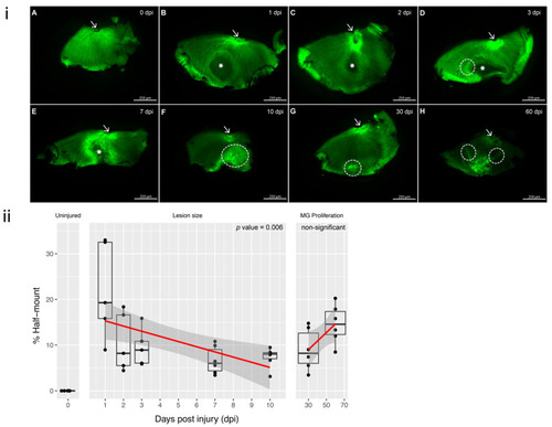
Figure 1. GFP retinal half-mounts showing the resolve of the lesion with the remaining scar; arrows point to the optic nerve for easier orientation: (i) (A) Uninjured (0 dpi) retinal half-mount with no lesion. (B,C) 1 and 2 dpi: lesion area is represented with a *, corresponding to a darker shaded area within the half-mount. (D) 3 dpi: the original circular lesion (*) has changed and there is a brighter margin surrounding the left side of the lesion (circled). (E) 7 dpi: reduced size of the lesion (*) and increased fluorescence within the margin. (F) 10 dpi: closing of the lesion with increased intensity of fluorescence, CMZ has been disrupted (circled). (G) 30 dpi: the lesion has resolved, and only reduced margin is visible near the CMZ (circled). (H) 60 dpi: the margin shows gathering of the tissue as the retina reaches the defect area, resulting in a fold at the site (circled). Each half-mount represents a separate animal. Scale bars represent 250 µm. (ii) Surrounding MG fluorescence intensity. The response to cryo-injury was quantified by calculating the percentages of the lesion area relative to the total area of the retinal half-mount. Linear regression was performed for (1) lesion size throughout 1–7 dpi and (2) MG proliferation between 10 and 60 dpi. A strong association for lesion size reducing from 1 to 10 dpi (p value = 0.00626). MG proliferation was notably increased from 10 dpi (p-value 0.0508, ns). Time points 1 dpi, 2 dpi and 3 dpi n = 5 independent animals and time points uninjured, 7 dpi, 10 dpi, 30 dpi and 60 dpi contain n = 6 independent animals.
|
Expression Data
Expression Detail
Antibody Labeling
Phenotype Data
Phenotype Detail
Acknowledgments
This image is the copyrighted work of the attributed author or publisher, and
ZFIN has permission only to display this image to its users.
Additional permissions should be obtained from the applicable author or publisher of the image.
Full text @ Cells