
FIGURE
Figure 4
- ID
- ZDB-FIG-220430-100
- Publication
- Yan et al., 2022 - Functional Study of TMEM163 Gene Variants Associated with Hypomyelination Leukodystrophy
- Other Figures
- All Figure Page
- Back to All Figure Page
Figure 4
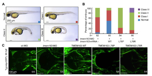
Figure 4. Functional analysis of TMEM163 mutations in zebrafish injected with translation-blocking morpholinos: (A) Representative images of the normal (blue), mild (red), severe (green) and profound (purple) phenotypes observed in the tmem163-MO, MO+TMEM163 WT, MO+TMEM163 L76P, MO+TMEM163 L76R injected groups at 48 hpf. Scale bar: 200 μm. (B) Percentage of living embryos showing any phenotype at 48 hpf. Co-injection of tmem163-MO with a human wild-type TMEM163 mRNA partially rescued phenotypes of tmem163 morphants such as degeneration of CNS, hydrocephalus, and bent tails, whereas injection of TMEM163 mRNA bearing the p.L76P or p.L76R mutation failed to rescue the phenotype. The number of embryos examined is listed under each bar. (C) The myelination in CNS is disrupted. Illustration of the Tg(mbp:GFP) larvae injected with control-MO, tmem163-MO, TMEM163 WT, TMEM163 L76P, TMEM163 L76R at 96 hpf (dorsal views with anterior to the left). Both the mutations disorganize the myelin in the brain. The ratios of affected embryos are indicated. Scale bar: 100 μm.
|
Expression Data
| Gene: | |
|---|---|
| Fish: | |
| Knockdown Reagents: | |
| Anatomical Term: | |
| Stage: | Day 4 |
Expression Detail
Antibody Labeling
Phenotype Data
| Fish: | |
|---|---|
| Knockdown Reagents: | |
| Observed In: | |
| Stage: | Day 4 |
Phenotype Detail
Acknowledgments
This image is the copyrighted work of the attributed author or publisher, and
ZFIN has permission only to display this image to its users.
Additional permissions should be obtained from the applicable author or publisher of the image.
Full text @ Cells