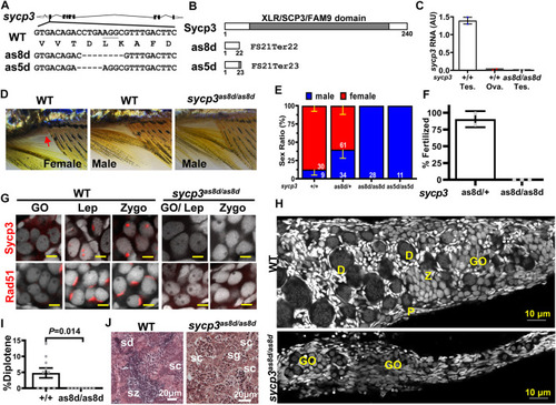
Disruption of sycp3 leads to infertile males with germ cell arrest during meiosis. (A). Maps of sycp3 and the sites of the mutations. The mutations are located in sycp3 exon 2. (B). The domain structures of wild type and mutant Sycp3. Both as8d and as5d mutations cause premature protein termination. (C). The RNA expression of sycp3 decreased in sycp3 mutant. (D)The external morphology of WT and sycp3 mutants. Females have a genital papilla (arrowhead) and yellowish fins. Males lack genital papilla and their fins are brownish. (E). The Sex Ratio of sycp3 mutants. The male is represented by the blue bar. Red bar represents females. The numbers of fish used in the analysis are shown inside the bars. (F). Fertilization test showing the frequency of 3-month male fish (genotype indicated below the X-axis) to produce offspring. (G). Immunofluorescence staining of 14-dpf WT and sycp3as8d/as8d gonads with Sycp3 and Rad51 antibodies (Wilson, High et al.). DAPI staining is shown in white color. The developmental stages of the meiotic germ cells are marked at the top of each figure. GO: gonocyte, Lep: leptotene, Zygo: zygotene. (H). The 1-month WT but not sycp3as8d/as8d gonad contain diplotene germ cells. D: diplotene, P: pachytene. (I). Quantitation of the % diplotene germ cells in a gonad. The p values were calculated using Student’s t test. (J). Testicular morphology showing sycp3as8d/as8d mutant germ cells do not go beyond spermatocytes (sc) while WT germ cells reach spermatozoa (sz). sg: spermatogonia, sd: spermatid.
|
