FIGURE SUMMARY
Title

Zebrafish Establish Female Germ Cell Identity by Advancing Cell Proliferation and Meiosis

Authors
Pan, Y.J., Tong, S.K., Hsu, C.W., Weng, J.H., Chung, B.C.
Source
Full text @ Front Cell Dev Biol

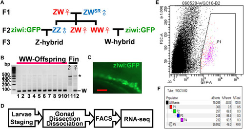
Isolation of germ cells from juvenile zebrafish with defined sex. (A). The flow chart for the generation of W- and Z-hybrids. Sex-reversed ZWSR Nadia males were crossed with ZW females to obtain the WW females in the F2 generation. The WW and ZZ fish were each mated with the Tg (ziwi: GFP) in the TL genetic background to obtain W- or Z-hybrid offspring with fluorescent germ cells. (B). Gel electrophoresis showing the detection of the W-fragment in the genome of 10 offsprings from the WW mother after PCR amplification (lanes 1–10). Lanes 11 and 12 are genomic DNAs from the fin of a male and a female of the Nadia strain as controls, respectively. The W fragment is 86 bps in length. The star marks nonspecific PCR bands. (C). The gonad of a Tg (ziwi: GFP) fish with fluorescent germ cells. (D). The flow chart for transcriptome analysis. Larvae were first staged to ensure proper development before their gonads were dissected. Their germ cells were then dissociated and sorted by FACS before RNA-seq analysis. (E). A representative FACS plot shows the selection of GFP-positive germ cells (P1). (F). A typical FACS data set showing the procurement of 232 GFP+ germ cells (pink group, 0.3% of all cells). Blue-group (P3) and pink-group (P4) cells were obtained after cell size quantity control. The green group (P2) selects for live cells that do not stain with propidium iodide. Black dots represent other cells and cell debris.

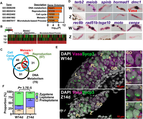
Meiosis as the hallmark of 14-dpf female germ cell transcriptome. (A). Gene ontology analysis of 1692 genes upregulated in the W-hybrid. (B). Expression of genes in the meiosis I, reproduction, cell cycle, and DNA metabolism at 10, 12, and 14 dpf. One gene is represented by a line, and the color represents the expression level normalized by Z-score. (C). A Venn diagram showing the overlaps of genes involved in meiosis I, reproduction, cell cycle, and DNA metabolism. (D). in situ hybridization of 10 meiotic genes in the 14-dpf W- and Z-hybrids. (E). The immunostaining of Sycp3 in gonocyte (GO), preleptotene (PL), leptotene (L), and zygotene (Z) germ cells. Germ cells marker Vasa and meiotic gene marker Sycp3 are magenta and green, respectively. DAPI are shown in white. The boxed regions are enlarged at the right side (1–4). (F). The W-hybrid advances meiosis faster than the Z-hybrid at 14 dpf. The proportion of preleptotene (yellowish), leptotene (blue), and zygotene (light green) are shown. The p value was calculated using the chi-squared analysis.

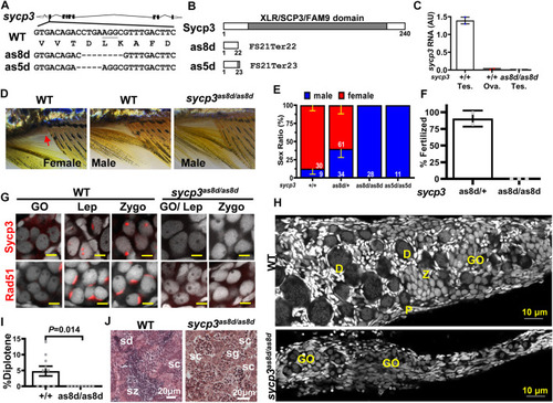
Disruption of sycp3 leads to infertile males with germ cell arrest during meiosis. (A). Maps of sycp3 and the sites of the mutations. The mutations are located in sycp3 exon 2. (B). The domain structures of wild type and mutant Sycp3. Both as8d and as5d mutations cause premature protein termination. (C). The RNA expression of sycp3 decreased in sycp3 mutant. (D)The external morphology of WT and sycp3 mutants. Females have a genital papilla (arrowhead) and yellowish fins. Males lack genital papilla and their fins are brownish. (E). The Sex Ratio of sycp3 mutants. The male is represented by the blue bar. Red bar represents females. The numbers of fish used in the analysis are shown inside the bars. (F). Fertilization test showing the frequency of 3-month male fish (genotype indicated below the X-axis) to produce offspring. (G). Immunofluorescence staining of 14-dpf WT and sycp3as8d/as8d gonads with Sycp3 and Rad51 antibodies (Wilson, High et al.). DAPI staining is shown in white color. The developmental stages of the meiotic germ cells are marked at the top of each figure. GO: gonocyte, Lep: leptotene, Zygo: zygotene. (H). The 1-month WT but not sycp3as8d/as8d gonad contain diplotene germ cells. D: diplotene, P: pachytene. (I). Quantitation of the % diplotene germ cells in a gonad. The p values were calculated using Student’s t test. (J). Testicular morphology showing sycp3as8d/as8d mutant germ cells do not go beyond spermatocytes (sc) while WT germ cells reach spermatozoa (sz). sg: spermatogonia, sd: spermatid.

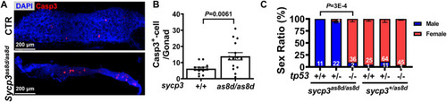
The sex reversal of sycp3 mutant fish is caused by germ cell apoptosis and can be compensated by additional tp53 mutation. (A). The immunostaining of apoptotic marker Caspase-3 (Wilson, High et al.). DAPI staining is blue for nuclei. (B). Quantification results of Caspase-3. Apoptotic germ cells are increased in the 1 month old sycp3as8d/as8d gonad. (C). The sex ratios of tp53-/-;sycp3as8d/as8d mutants. While sycp3as8d/as8d fish are all males, many females have the tp53-/-;sycp3as8d/as8d genotype. Blue bar represents males and the red bar represents females.

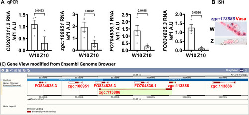
Genes clustered on chromosome 17 are upregulated in 10-dpf germ cells of the W-hybrid. (A). Higher expression of CU207311.2, zgc:100951, FO704836.1, FO834825.3, in the W-hybrid than the Z-hybrid at 10 dpf as detected by RT-PCR. The Y-axis is the RNA level in arbitrary units (A.U.) after normalization with an internal control (ef1). (B). The expression of zgc:113886 (blue-purple) is higher in the W-hybrid shown by in situ hybridization. Vasa antibody stains germ cells red in Alexa fluor 546. (C). All these upregulated genes are clustered in the same locus on chromosome 17 as shown by the Ensembl Gene view.

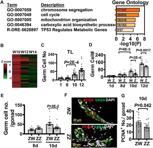
Transcriptome analysis of 10-dpf germ cells identifies the advanced germ cell proliferation as a sign of zebrafish female differentiation. (A). Gene ontology analysis of 668 differentially expressed genes (DEGs) upregulated in the 10 dpf W-hybrid. (B). The heat map of DEGs in the pathways of chromosome segregation (GO:0007059) and cell cycle (GO:0007049) are high in the W-hybrid at 10 dpf and gradually decreased at 12 and 14 dpf. The expression level is normalized by a z-score (C). The germ cell numbers of TL laboratory strain at 1, 8, 10, and 12 dpf. Germ cells significantly proliferate at 10 dpf. (D). The germ cell numbers of W- and Z-hybrid become different from 10 dpf on. (E). Germ cell number in ZW genotype of Nadia is higher than that in ZZ gonad at 10 dpf. (F). Double immunofluorescence showing PCNA staining for proliferating germ cell and Vasa (green) for germ cells. DAPI staining is white for nuclei. (G). Quantitation of PCNA+-germ cells, showing ZW gonads contain more proliferating germ cells than ZZ gonads at 10 dpf. One dot represents data from one gonad. The two-tailed and unpaired student t-test used for statistical analysis.

Acknowledgments
This image is the copyrighted work of the attributed author or publisher, and ZFIN has permission only to display this image to its users. Additional permissions should be obtained from the applicable author or publisher of the image. Full text @ Front Cell Dev Biol