|
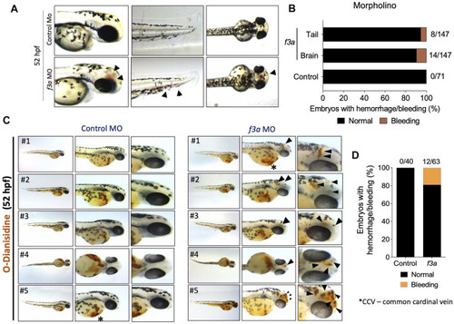
f3a knockdown shows bleeding phenotype: (A) Lateral and dorsal views of head, and lateral view of tail in live embryos. The arrows depict brain and tail bleeding at 52 hpf and (B) quantification of the same. Numbers in the bars represent the ratios used to calculate the percentages. (C) Bright-field images of zebrafish embryos stained with O-dianisidine (OD) and imaged at 48 hpf. Black arrows indicate sites where abnormal accumulation of hemoglobinized blood was detected in the head region (control MO and f3a MO). (D) Percentage of cerebral hemorrhage in embryos at 52 hpf. Numbers in the bars represent the ratios used to calculate the percentages. Data were pooled from three independent experiments.
|