FIGURE

FIGURE 1

ID
ZDB-FIG-220406-48
Publication
Schwarzer et al., 2022 - Reactivation of the Neurogenic Niche in the Adult Zebrafish Statoacoustic Ganglion Following a Mechanical Lesion
Other Figures
All Figure Page
Back to All Figure Page
FIGURE 1

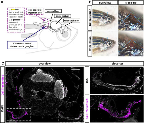
Establishment of a lesion paradigm of the adult zebrafish statoacoustic ganglion (SAG). (A) Scheme of the zebrafish head with the brain inside showing the injection position in relation to the brain and the SAG. A 20-gauge needle was used to open the skull before inserting a Hamilton syringe vertically approximately 1.2- to 1.5-mm deep into the skull along the dorsal–ventral axis. Subsequently, the Hamilton syringe was either removed (sham-treated control) or 5–8 µl of either 0.9% NaCl or 100 µM ouabain solution was slowly injected. (B) Overview of live adult zebrafish before and after injection. Close-ups of the blue dotted boxes show the injection side within the triangular bone (dotted blue line). Lower close-up shows the hole after injection (dotted yellow circle). (C) Immunohistochemistry of a transverse cross section at the height of the anterior SAG following CellTracker Red injection into the right inner ear showed a strong labeling of the SAG on the injected side but no labeling of the unlesioned side (ULS). Scale bar: 200 µm.

Expression Data

Expression Detail
Antibody Labeling
Phenotype Data

Phenotype Detail
Acknowledgments
This image is the copyrighted work of the attributed author or publisher, and ZFIN has permission only to display this image to its users. Additional permissions should be obtained from the applicable author or publisher of the image. Full text @ Front Cell Dev Biol