
FIGURE
Fig 3
- ID
- ZDB-FIG-220319-52
- Publication
- Liu et al., 2022 - Rod genesis driven by mafba in an nrl knockout zebrafish model with altered photoreceptor composition and progressive retinal degeneration
- Other Figures
- All Figure Page
- Back to All Figure Page
Fig 3
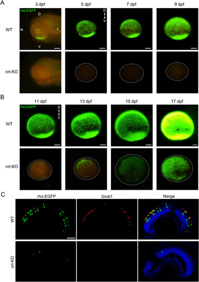
Rods were labeled with EGFP by crossing the WT or nrl-KO zebrafish with the Tg(rho:EGFP) transgenic line. Fluorescence was observed every 2 days from 3 dpf. Representative images of WT and nrl-KO retinas are shown in (A) for 3–9 dpf and (B) for 11–17 dpf. The dotted circles indicate the boundaries of the retinas. D, dorsal; V, ventral; N, nasal; T, temporal. Scale bars: 100 μm. (C) Immunostaining of retinal sections for Gnat1 (the rod transducin alpha-subunit) showed exact co-localization with EGFP in rods at 7 dpf. Rods were barely observed in the nrl-KO retinas. Scale bar: 50 μm. |
Expression Data
| Gene: | |
|---|---|
| Antibody: | |
| Fish: | |
| Anatomical Term: | |
| Stage Range: | Protruding-mouth to Days 14-20 |
Expression Detail
Antibody Labeling
Phenotype Data
| Fish: | |
|---|---|
| Observed In: | |
| Stage Range: | Pec-fin to Days 14-20 |
Phenotype Detail
Acknowledgments
This image is the copyrighted work of the attributed author or publisher, and
ZFIN has permission only to display this image to its users.
Additional permissions should be obtained from the applicable author or publisher of the image.
Full text @ PLoS Genet.