|
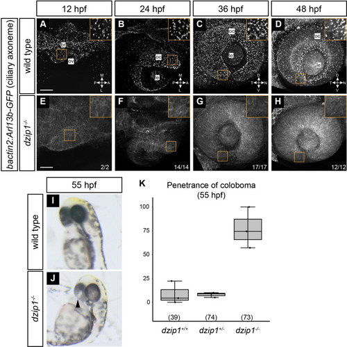
<italic toggle='yes'>dzip1</italic> mutants have no cilia in the eye and display coloboma.(A-H) Wild type (A-D) and dzip1ts294e mutants (E-H) visualized for cilia (Tg(bactin2:Arl13b-GFP)) at 12 hpf (A, E), 24 hpf (B, F), 36 hpf (C, G), and 48 hpf (D, H). A, B, E, and F are dorsal views of 3-dimensional renderings; while C, D, G, and H are lateral views of 3-dimensional renderings. (A-D) GFP-positive cilia are visualized as distinct puncta present throughout the lens, eye, and embryo midline (A, magenta arrowhead). (E-H) Cilia are completely absent in dzip1-- embryos. n (fraction of dzip1-/- embryos showing complete absence of cilia) is shown at the bottom right of each panel. Insets show zoomed views for each timepoint and genotype. (I-K) dzip1-/- mutants display coloboma. (I) Wildtype embryo at ~55 hpf; the eye is evenly pigmented; there is no coloboma. (J) dzip1ts294e mutant embryo, ~55 hpf; coloboma is visible as a hypopigmented region at the back of the eye (arrowhead). (K) Penetrance of the coloboma phenotype at 55 hpf. n (embryos) for each genotype shown at the base of the graph. Scale bar: 50 μm. M, medial; L, lateral; A, anterior; P, posterior; D, dorsal; V, ventral; N, nasal; T, temporal; le, lens; oc, optic cup.
|