
Fig. 2
- ID
- ZDB-FIG-220308-10
- Publication
- Braunstein et al., 2021 - Basal epidermis collective migration and local sonic hedgehog signaling promote skeletal branching morphogenesis in zebrafish fins
- Other Figures
- All Figure Page
- Back to All Figure Page
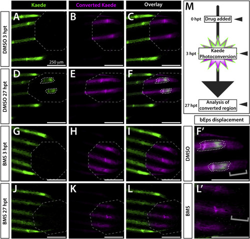
Active Shh/Smo signaling is restricted to a narrow distal stretch of each developing fin ray. (A–L) Whole mount fluorescence images of the distal aspect of the caudal fin of 25 dpf ptch2:Kaede fish treated with DMSO (A–F) or BMS-833923 (BMS; G-L). Images are from the time of Kaede photoconversion from green to red (false colored magenta) fluorescence (3 h post treatment (hpt); A-C, G-I) and 24 h later (27 hpt, D-F, J-L). Grey dashed octagons mark photoconverted regions of interest (ROIs). The 3 hpt overlay (C) demonstrates complete Kaede photoconversion. The same fish at 27 hpt displays a small patch of newly produced green Kaede (white dotted outlines in D, F) within the ROI. Slight splitting of the new green Kaede domain indicates the onset of ray branching. The BMS-treated fish shows no new Kaede within the ROI (J–L). (F’, L’) Zoom view of distal ray regions. Grey brackets mark presence or absence of distally displaced basal epidermis retaining photoconverted Kaede. The few green Kaede+basal epidermal cells in (J, L) moved into the photoconverted region without producing new Kaede. (M) Schematic of the time course for drug treatment, photoconversion, and imaging. Imaged fish represent groups of n = 8. Scale bars are 250 μm. |
Reprinted from Developmental Biology, 477, Braunstein, J.A., Robbins, A.E., Stewart, S., Stankunas, K., Basal epidermis collective migration and local sonic hedgehog signaling promote skeletal branching morphogenesis in zebrafish fins, 177-190, Copyright (2021) with permission from Elsevier. Full text @ Dev. Biol.