FIGURE

FIGURE 5

ID
ZDB-FIG-220305-14
Publication
Huang et al., 2022 - An In Vivo Fluorescence Resonance Energy Transfer-Based Imaging Platform for Targeted Drug Discovery and Cancer Therapy
Other Figures
All Figure Page
Back to All Figure Page
FIGURE 5

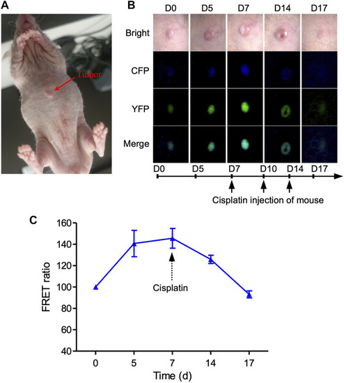
Imaging of cell apoptosis in xenograft tumor mouse in vivo. (A) Imaging of 231-C3 xenograft nude mice and FRET channels of a tumor with no drug treatment. 100 µL 5×10⁶/ml 231-C3 cells were injected into each site of nude mice. Mice were injected with 6 mg/kg cisplatin after the establishment of the tumor xenograft. (B) Imaging of 231-C3 xenograft tumor nude mice with cisplatin treatment. (C) Quantification of FRET ratio in xenograft nude mice tumors after 6 mg/kg cisplatin treatment. In vivo imaging of mouse tumor was performed using a Leica fluorescent microscope. CFP (ex: 440 nm; em: 480 nm), YFP (ex: 440 nm; em: 535 nm). Data are expressed as mean ± s.e.m. n = 3.

Expression Data

Expression Detail
Antibody Labeling
Phenotype Data

Phenotype Detail
Acknowledgments
This image is the copyrighted work of the attributed author or publisher, and ZFIN has permission only to display this image to its users. Additional permissions should be obtained from the applicable author or publisher of the image. Full text @ Front Bioeng Biotechnol