
Figure 4
- ID
- ZDB-FIG-220219-69
- Publication
- Codolo et al., 2022 - Macrophage-Mediated Melanoma Reduction after HP-NAP Treatment in a Zebrafish Xenograft Model
- Other Figures
- All Figure Page
- Back to All Figure Page
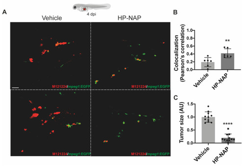
HP-NAP treatment favors the interaction between tumor cells and macrophages and early affects tumor size. Tg(mpeg1:EGFP)gl22 zebrafish embryos were xenotransplanted with M121224 melanoma cells (red), injected or not with HP-NAP at 2 dpi and observed at 4 dpi. (A) Representative 2D projections of confocal single plane images of the yolk-sac region of embryos at 4 dpi. Magnification: 40×. Scale bar: 20 µm. (B) Scatter plots show the colocalization (Pearson’s correlation) between green (macrophages) and red (tumor cells) signals in vehicle- and HP-NAP-injected fishes. At least 5 Regions of Interest (ROIs) per sample of 3 independent experiments were analyzed. Values are shown as mean ± SEM and analyzed by Student’s t test; **, p < 0.01; n = 6 for each condition. (C) Scatter plots show the quantification of the tumor size (AU: Arbitrary Unit) at 4 dpi. Values are expressed as mean ± SEM and analyzed by Student’s t test; ****, p < 0.0001; n = 11 for each condition, from 3 independent experiments. |