
Figure 1
- ID
- ZDB-FIG-220212-9
- Publication
- Okamoto et al., 2022 - Ca2+-imaging and photo-manipulation of the simple gut of zebrafish larvae in vivo
- Other Figures
- All Figure Page
- Back to All Figure Page
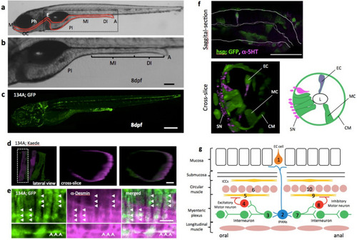
Simple structure of the zebrafish gut. (a) Lateral view of zebrafish larva at 8 dpf, indicating the principal digestive tract. The intestine is divided into three parts: proximal intestine (PI), middle intestine (MI), and the distal part of the intestine (DI)10. The PI is also referred to as the intestinal bulb and is thought to have similar functions as the stomach, which zebrafish do not have. The MI is thought to absorb nutrients and plays a role in mucosal immunity. The DI is analogous to the colon. M mouth, OC oral cavity, Ph pharynx, E esophagus, PI proximal intestine, MI middle intestine, DI distal intestine, A anus. (b) A higher-magnification image of the area indicated by the square in (a). Scale bar, 50 μm. (c) Lateral view of SAGFF(LF)134A; Tg(UAS: GFP) at 8 dpf. Scale bar, 200 μm. (d) Live confocal image of individual circular smooth muscles visualized by green to red photoconversion of Kaede fluorescence in SAGFF(LF)134A; Tg(UAS: Kaede) at 5 dpf. Lateral projection view shows two muscles photoconverted. Left side view. Cross-slice view of the dotted area shows a single red muscle. Scale bar, 10 μm. (e) Saggital sections of a confocal image of SAGFF(LF)134A; Tg(UAS: GFP) at 8 dpf stained with anti-Desmin antibody. Upper panels demonstrate Desmin filaments (in magenta, shown by triangles) located inside of each circular smooth muscles expressing GFP. Lower panels show an example of occasional appearance of longitudinal muscle cell expressing GFP, containing Desmin filaments (arrows). Scale bar, 10 μm. (f) Saggital section and cross-slice of the gut, indicating a variety of cell types visualized at 5 dpf in Tg(hsp70: Gal4); Tg(UAS: GFP) by heat shock at 3 dpf. Double immunostaining for anti-GFP (green) and anti-5-HT (magenta). EC serotonergic endochromaffin cells, MC mucosal cells, L lumen, CM circular muscle, SN neurites of serotonergic neurons. Scale bar, 50 μm. (g) Schematic diagram showing the hypothetical circuitry for the peristaltic reflex in the zebrafish gut. 1, endochromaffin cell; 2, intrinsic primary afferent cell; 3, ascending interneuron; 4, excitatory motoneuron; 5, 9, interstitial cells of Cajal; 6, 10, circular smooth muscles; 7, descending interneuron; 8, inhibitory motoneuron. |