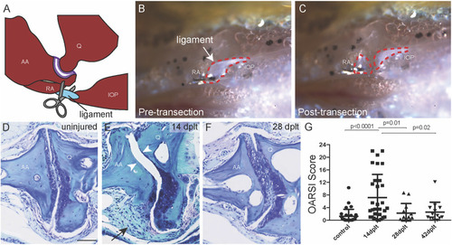
Joint degeneration and recovery following ligament transection surgery. (A) Schematic of the bones of the zebrafish jaw relative to the jaw joint (purple) and the interopercular–mandibular (IOM) ligament (blue) that is surgically transected. (B,C) Reflected light images of the zebrafish jaw in ventral view show the IOM ligament (dotted red outline) before (B) and immediately after (C) transection. (D–F) Toluidine blue staining of uninjured adult jaw joint (D) and IOM-transected jaw joint at 14 days post-ligament transection (dplt) (E) and 28 dplt (F). The arrowheads point to the area of cartilage erosion and the arrow to the synovial hyperplasia. (G) Quantification of jaw joint degeneration after IOM transection using the zebrafish OARSI scoring method; n = 29 uninjured; n = 31 at 14 dplt; n = 13 at 28 dplt; n = 14 at 42 dplt. Individual data points, mean, and standard error of the mean are shown. The p-values are calculated by ANOVA and Tukey’s multiple-comparison test. AA, anguloarticular bone; IOP, interopercular bone; Q, quadrate bone; RA, retroarticular bone. Scale bar = 50 μm.
|
