
FIGURE
Figure 3
- ID
- ZDB-FIG-220131-533
- Publication
- Lu et al., 2022 - Effect of Lipopolysaccharides on Liver Tumor Metastasis of twist1a/krasV12 Double Transgenic Zebrafish
- Other Figures
- All Figure Page
- Back to All Figure Page
Figure 3
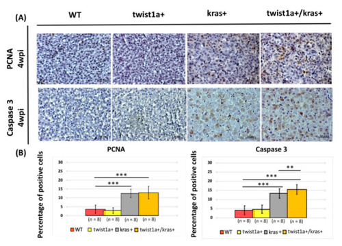
Main hallmarks of cell proliferation and cell apoptosis in liver tissues from twist1a+/kras+ double transgenic zebrafish. (A) Immunohistochemical staining micrograph showing proliferating and apoptotic cells in liver cross-sections from wild-type, twist1a+, kras+, and twist1a+/kras zebrafish. (B) Quantification of the percentage of positive cells for the all fields following PCNA and caspase-3 staining at 4 wpi using ImageJ. Scale bar: 50 μm. Student’s t-tests were used to assess differences between variables: ** p < 0.01, *** p < 0.001. |
Expression Data
Expression Detail
Antibody Labeling
Phenotype Data
| Fish: | |
|---|---|
| Conditions: | |
| Observed In: | |
| Stage: | Adult |
Phenotype Detail
Acknowledgments
This image is the copyrighted work of the attributed author or publisher, and
ZFIN has permission only to display this image to its users.
Additional permissions should be obtained from the applicable author or publisher of the image.
Full text @ Biomedicines