
Fig. 3
- ID
- ZDB-FIG-220113-2
- Publication
- Mullapudi et al., 2019 - Disruption of the pancreatic vasculature in zebrafish affects islet architecture and function
- Other Figures
- All Figure Page
- Back to All Figure Page
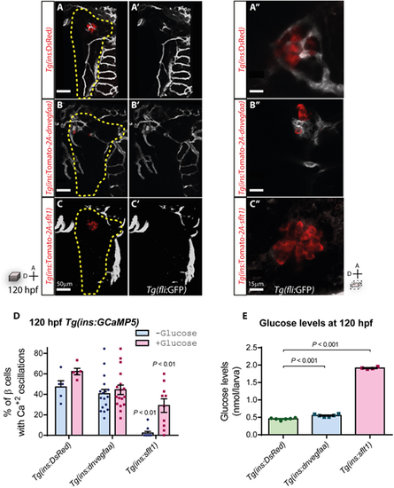
sFlt1 expression in β-cells severely impairs their function. (A-C) Confocal projection images of the vasculature (white) in 120 hpf Tg(fli:GFP) larvae, in combination with the indicated transgenic line. Yellow dashed lines outline the pancreas. A′-C′ show the endothelial cells. (A″-C″) Confocal planes of β-cells (red) and the surrounding vasculature (white) in 120 hpf Tg(fli:GFP) larvae, in combination with the indicated transgenic line. (D) Percentage of β-cells responsive to physiological (−Glucose) and exogenous (+Glucose) glucose, measured as visual fluctuations in fluorescence in 120 hpf Tg(ins:GCaMP5) larvae, n=5-17 animals. (E) Glucose levels in 120 hpf animals, n=4-6 replicates, 10 animals per replicate. Data are mean±s.e.m; individual data points are shown. P-values from t-tests are presented. A, anterior; D, dorsal. |