|
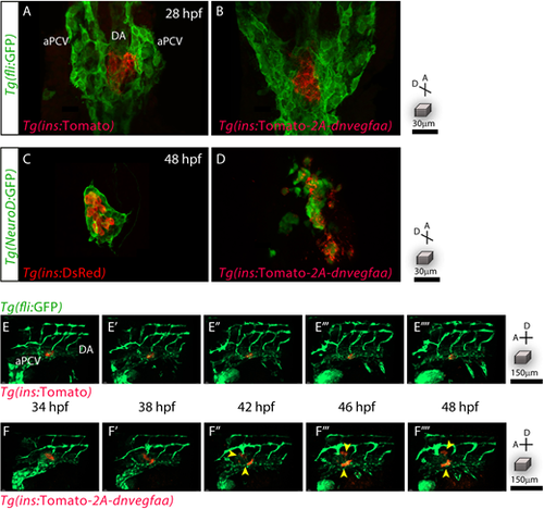
β-Cell-specific dnVegfaa expression disrupts pancreatic islet architecture. (A,B) Pancreatic β-cells (red) and the endothelial cells (green) in 28 hpf Tg(ins:Tomato); Tg(fli:GFP) and Tg(fli:GFP); Tg(ins:dnvegfaa) embryos, viewed ventrally. (C,D) Pancreatic endocrine cells (green) and β-cells (red) in 48 hpf Tg(NeuroD:GFP); Tg(ins:DsRed) and Tg(NeuroD:GFP); Tg(ins:dnvegfaa) embryos, viewed ventrally. (E-F) Confocal projection images of Tg(ins:Tomato); Tg(fli:GFP) and Tg(ins:dnvegfaa); Tg(fli:GFP) embryos from 34 to 48 hpf, viewed laterally. Yellow arrowheads point to dispersing β-cells. Maximum intensity projections are presented. A, anterior; D, dorsal.
|