
Fig. 4.
- ID
- ZDB-FIG-211216-239
- Publication
- Miles et al., 2021 - Usher syndrome Type 1-associated gene, pcdh15b, is required for photoreceptor structural integrity in zebrafish
- Other Figures
- All Figure Page
- Back to All Figure Page
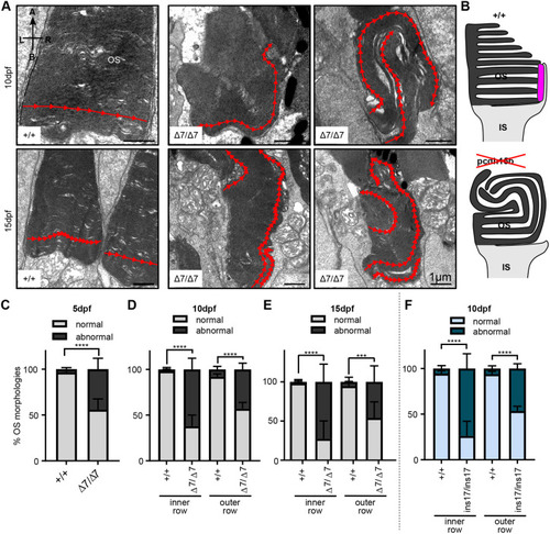
Ultrastructure of OSs in pcdh15b mutant photoreceptors exhibit abnormal directional growth of discs. (A) Representative TEM images of OSs of wild-type siblings and Δ7 mutants at 10 dpf and 15 dpf. Red arrow line shows disc direction. Directional arrows at the top left of the first image indicate positioning. (B) Schematic of the effects of pcdh15b mutation on the growth of the OS discs. Pcdh15 is shown in +/+ by the magenta-coloured shape. (C-F) Quantification of the percentage of ‘normal’ and abnormal OSs in wild-type siblings and pcdh15b mutants at different time points and mutant types (Δ7 and ins17). Statistical analyses in C-F were performed using Student's t-tests (unpaired, two-tailed). ***P≤0.001, ****P≤0.0001. OSs were analysed across the entire ONL for the 5 dpf and 10 dpf data, and the central retina for 15 dpf data. Number of eyes/OSs analysed: (B) 5 dpf: +/+, 10 eyes/2123 OSs; Δ7/Δ7, 10 eyes/1874 OSs; (C) 10 dpf: +/+, 9 eyes/1990 OSs; Δ7/Δ7, 12 eyes/2457 OSs; (D) 15 dpf: +/+, 6 eyes/320 OSs; Δ7/Δ7, 6 eyes/291 OSs; (E) 10 dpf: +/+, 8 eyes/1260 OSs; ins17/ins17, 7 eyes/956 OSs. A, apical; B, basal; IS, inner segment; L, left; ONL, outer nuclear layer; OS, outer segment; R, right. |
| Fish: | |
|---|---|
| Observed In: | |
| Stage Range: | Day 5 to Days 14-20 |