
Fig. 3.
- ID
- ZDB-FIG-211118-102
- Publication
- Ren et al., 2021 - Novel zebrafish polycystic kidney disease (PKD) models reveal functions of the Hippo pathway in renal cystogenesis
- Other Figures
- All Figure Page
- Back to All Figure Page
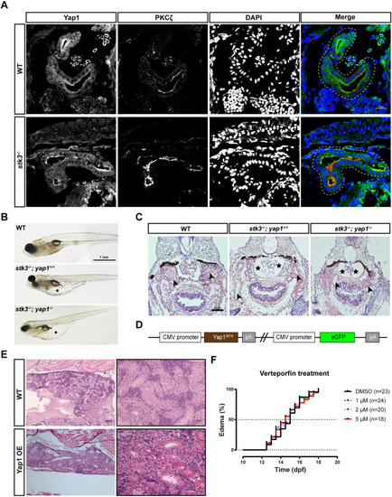
Functional analysis of Yap1 in zebrafish stk3−/− mutants. (A) Immunofluorescent staining of Yap1 and PKCζ in cryostat sections of stk3−/− larvae and WT siblings at 12 dpf. DAPI stains nuclei. The merged images are presented with Yap1 staining in green, PKCζ in red and DAPI in blue. Dashed line loops indicate the renal tubules. Scale bar: 20 μm. (B) Morphology of larvae at 15 dpf. Asterisks indicate the edema. Scale bar: 1 mm. (C) H&E staining of paraffin sections of larvae at 15 dpf. stk3−/−;yap1−/− double mutants exhibited the same phenotype as stk3−/− single mutants. Enlarged Bowman's space (asterisks) and pronephric tubule (arrowheads) dilation could be detected. Scale bar: 50 μm. (D) Schematic diagram of the expression plasmid for Yap1 OE fish line. (E) H&E staining of paraffin sections of adult kidneys at 90 dpf. No obvious difference between Yap1 OE fish and WT controls could be detected. Scale bars: 100 μm (left) and 20 μm (right). (F) Kaplan–Meier plots for the onset of edema in stk3−/− larvae under verteporfin treatment. Treatment with a different dose of verteporfin (1, 2 and 5 μM) did not influence the onset of edema compared to DMSO control. Analysis with log-rank (Mantel–Cox) test showed no statistical significance. |