
Fig. 3
- ID
- ZDB-FIG-211105-28
- Publication
- Hatzold et al., 2021 - The Kunitz-type serine protease inhibitor Spint2 is required for cellular cohesion, coordinated cell migration and cell survival during zebrafish hatching gland development
- Other Figures
- All Figure Page
- Back to All Figure Page
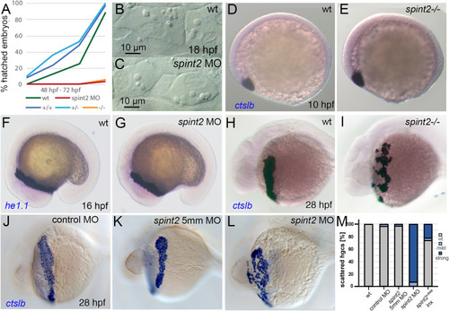
A. Percentage of hatched embryos during 48–72 hpf, n = 24–50. B–C. DIC images of hatching gland cells displaying characteristic granules in wild type (B) and spint2 MO (C) at 18 hpf. D-G. WISH of hatching gland marker genes shows unaltered expression of ctslb in wild type (D) and spint2 mutant (E) at end of gastrulation (10 hpf), and of he1.1 in wild type (F) and spint2 morphant (G) at 16 hpf. H-L. WISH of ctslb at 28 hpf showing hatching gland cells that are organized in a belt-like structure in wild type sibling (H), standard control- (J) or 5 mm control morpholino-injected (K) embryo, but disorganized and scattered in spint2 mutant (I) and spint2 morphant (L). M. Quantification of the hatching gland cell phenotype of mutants and morphants shown in H – L. N = 3 independent experiments, n = 53–73 embryos. |
| Genes: | |
|---|---|
| Fish: | |
| Knockdown Reagents: | |
| Anatomical Terms: | |
| Stage Range: | Bud to Prim-5 |
| Fish: | |
|---|---|
| Knockdown Reagents: | |
| Observed In: | |
| Stage Range: | Bud to Prim-5 |
Reprinted from Developmental Biology, 476, Hatzold, J., Wessendorf, H., Pogoda, H.M., Bloch, W., Hammerschmidt, M., The Kunitz-type serine protease inhibitor Spint2 is required for cellular cohesion, coordinated cell migration and cell survival during zebrafish hatching gland development, 148-170, Copyright (2021) with permission from Elsevier. Full text @ Dev. Biol.