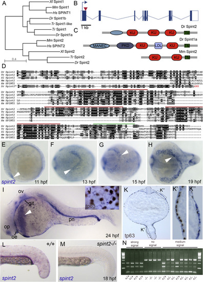
A. Phylogenetic tree of protein sequences of zebrafish (Dr), pufferfish (Takifugu rubripes, Tr), clawed frog (Xenopus laevis, Xl), mouse (Mm), and human (Hs) Spint1 and Spint2 proteins. The tree was created using COBALT and the Neigbor Joining method on the NCBI webpage. B. Schematic representation of zebrafish spint2 gene structure. Exons are indicated by rectangles, with coding regions in solid blue, introns are indicated with lines. The arrow head indicates the position targeted by the Crispr guide RNA. C. Schematic representation of Zebrafish Spint2 and Spint1a as well as mouse Spint2 protein domains. KU, Kunitz-type domain; PKD, polycystic kidney disease-like domain; MANEC, motif at N-terminus with eight-cysteines; TM, transmembrane domain. D. Multiple alignment of zebrafish (Dr), pufferfish (Takifugu rubripes, Tr), clawed frog (Xenopus laevis, Xl), mouse (Mm), and human (Hs) Spint2 protein sequences by Clustal Omega. Identical amino acid (aa) residues are indicated by black boxes, similar aa residues by grey boxes. Red lines indicate Kunitz-type domains, green line the transmembrane domain. E.-I. Whole mount in situ hybridization (WISH) with a spint2 probe shows expression in the epidermis and hatching gland cells throughout somitogenesis (E-H) and additional expression in the olfactory epithelium (olf), otic vesicle (ot), and pronephric duct (pd) at 24 hpf (I). J-K. WISH for spint2 at 24 hpf followed by tp63 immunostaining shows expression of spint2 in peridermal cells but not in tp63-positive basal cells in whole mounts (J) and cross sections (K, K′) as well as expression in hatching gland cells (K″). L,M. spint2 WISH in spint2+/+ and −/− embryos at 24 hpf. spint2 signal is not detectable in spint2fr49/fr49 embryos. N. Ethidumbromide agarose gel with genotyping results of embryos obtained from an incross of spint2+/fr49 parents after they had been stained via spint2 mRNA WISH. The obtained staining intensity of the individual embryos is indicated.
|
