|
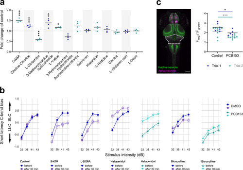
Involvement of neurotransmitters in startle latency.a Neurotransmitter levels in 6 dpf larvae exposed to 1000 nM PCB153 in comparison to control (n = 4, pool of 18 larvae per sample). Unpaired t-test GABA ***p = 0.00007, choline-chloride ***p = 0.0001, L-glutamine ***p = 0.0010, 3-methoxytyramine hydrochloride **p = 0.0089, L-valine *p = 0.0202. b Startle bias shift induced by 30-min exposure to serotonin, dopamine, and GABA neurotransmitter modulators in 6 dpf control and 100 nM PCB153 exposed larvae. Control (n = 172), 5-HTP (n = 80), L-DOPA (n = 94), haloperidol (Control n = 178, PCB153 n = 81), and bicuculline (Control n = 160, PCB153 n = 173). All data points are biologically independent samples from at least three independent experiments and mean ± SEM are shown in the plots. Significant differences to respective controls (GLM, p < 0.01) are indicated by open symbols. c Representative maximum intensity z-projection from confocal stack after photoconversions of freely swimming 6 dpf CaMPARI larvae. Scale bar = 100 µm. Ratio of red to green fluorescence intensity in the rostral hindbrain (indicated by dashed line in image) in DMSO (n = 14) and 1000 nM PCB153 (n = 14) exposed swimming larvae in 4 ˚C (cold) medium. Asterisks indicate significant differences to controls (two-way ANOVA, *p = 0.0277, ***p = 0.0004). All data points are biologically independent samples from two independent experiments (trials) and the horizontal lines are indicating the mean.
|