|
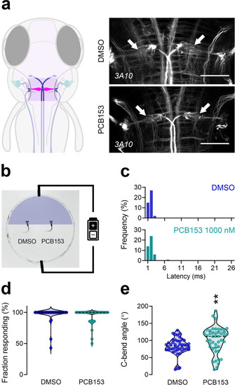
Effect of PCB153 on presence and function of the Mauthner cells.a Diagram indicating localization of Mauthner cells in the hindbrain and brain tissue immunostaining with anti-neurofilament 3A10 antibody labeling reticulospinal neurons in control and PCB153 treated larvae at 6 dpf (ventral view). Mauthner (arrow) cells were present in all brain tissues (DMSO: n = 23, PCB153: n = 21, biologically independent samples). Scale bar = 50 µm. b Diagram of electrical stimulation performed to assess the functionality of Mauthner cells. Both DMSO (n = 44) and PCB153 (n = 45) treated larvae show similar c latency to electrical stimulation and d response frequency, indicating that the Mauthner cells are present and functional, e while the C-bend angle in PCB153 exposed larvae was increased (Welch’s t-test, **p = 0.0091). All data points are biologically independent samples (one value representing the mean of seven consecutive electric field pulses) from three independent experiments and mean ± SD are shown in the plots. Mann–Whitney test was used for statistical analysis in (c), binomial GLM in (d), and unpaired two-tailed t-test with Welch’s correction in (e). Dashed lines represent median and quartiles and asterisks indicate significant differences to controls.
|