
Fig. 1
- ID
- ZDB-FIG-211012-1
- Publication
- Rattka et al., 2021 - Spen deficiency interferes with Connexin 43 expression and leads to heart failure in zebrafish
- Other Figures
- All Figure Page
- Back to All Figure Page
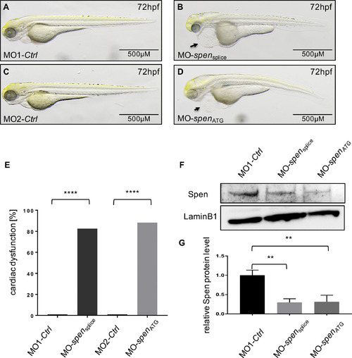
Lateral view of Spen-deficient (B,D) and control-injected embryos (A,C) at 72 h post fertilization (hpf). Spen deficiency results in development of a large pericardial edema at 72 hpf (B,D black arrow), while the controls don't display cardiac abnormalities (A,C). (E) 86% (190/221) MO-spensplice- and 79% (145/184) MO-spenATG- injected embryos display the same cardiac phenotype. Embryos with pericardial edema, bradycardia, and impaired contractility were counted as affected. (F) Efficacy of morpholino-mediated knockdown experiments was confirmed by Western Blot analysis; representative immunoblots are shown. (G) Relative quantification of Spen protein levels (n = 3). Images (A,B,C,D,F) have been cropped. Assessment of significance by chi-squared test (E) or t-test (G). *p < 0.05, **p < 0.01, ***p < 0.001, ****p < 0.0001. |
| Fish: | |
|---|---|
| Knockdown Reagents: | |
| Observed In: | |
| Stage: | Protruding-mouth |
Reprinted from Journal of Molecular and Cellular Cardiology, 155, Rattka, M., Westphal, S., Gahr, B.M., Just, S., Rottbauer, W., Spen deficiency interferes with Connexin 43 expression and leads to heart failure in zebrafish, 25-35, Copyright (2021) with permission from Elsevier. Full text @ J. Mol. Cell. Cardiol.