|
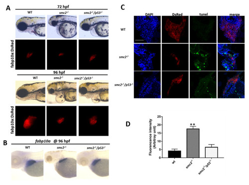
Activation of the p53-dependent apoptotic pathway contributed to the small liver phenotype in SMC2−/− mutants. (A) Phenotype comparison of WT, SMC2−/− and SMC2−/−/p53−/− embryos under the Tg (fabp10a:dsRed;ela3l:EGFP) transgenic background at 72 hpf and 96 hpf. (B) WT embryos, SMC2−/− and SMC2−/−/p53−/− embryos stained with the fabp10a probe at 96 hpf. (C) TUNEL analysis of apoptotic cells in the liver of SMC2−/−/p53−/− mutants compared to WT and SMC2−/− mutants at 96 hpf under the Tg (fabp10a:dsRed;ela3l:EGFP) transgenic background. Scale bar, 50 μm. (D) Quantitative analysis of the apoptotic cells in the liver. Fluorescence intensities of three WT embryos, three SMC2−/− mutant embryos and three SMC2−/−/p53−/− embryo across the liver were determined using the ImageJ software. **, p < 0.01.
|