FIGURE

Figure 9

ID
ZDB-FIG-211002-10
Publication
Chebli et al., 2021 - The localization of amyloid precursor protein to ependymal cilia in vertebrates and its role in ciliogenesis and brain development in zebrafish
Other Figures
All Figure Page
Back to All Figure Page
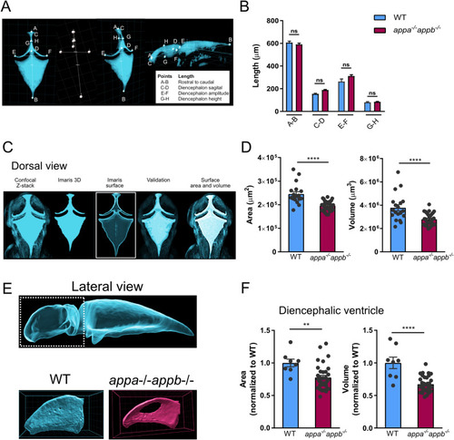
Figure 9

The appa−/−appb−/− 2 dpf larvae zebrafish exhibit smaller brain ventricle. Dorsal 3D surface rending of confocal stacks

taken from brain ventricles of dextran injected 2 dpf zebrafish larvae (A). Quantification of total ventricle surface area and volume show that both are decreased in appa-/-appb-/- larvae (B). Lateral 3D surface rending of confocal stacks from brain ventricles of dextran injected 2 dpf zebrafish larvae with close up on diencephalic ventricle (C). Quantification of surface area and volume of the diencephalic ventricle in WT and appa-/-appb-/- larvae (D). Measurement of gross ventricle morphology at 2dpf WT and appa-/-appb-/- larvae as the length (E). Distance between rostral to caudal, diencephalon ventricle sagittal length, amplitude and height show no significant difference in mutants (F). Data are reported as mean ± SEM. ** ρ < 0.01, **** ρ < 0.001. n: (B) WT = 19, appa-/-appb-/- = 34, (D) WT = 8, appa-/-appb-/- = 34, (F) WT = 5, appa-/-appb-/- = 4.

Expression Data

Expression Detail
Antibody Labeling
Phenotype Data
Fish:
Observed In:
Stage: Long-pec

Phenotype Detail
Acknowledgments
This image is the copyrighted work of the attributed author or publisher, and ZFIN has permission only to display this image to its users. Additional permissions should be obtained from the applicable author or publisher of the image. Full text @ Sci. Rep.