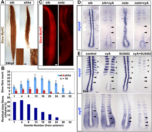
(A) Immunodetection showing slow fibre reduction is greater in tail than trunk in shha mutants. Insets show individual fibres magnified. (B) Quantification of experiment in A. Upper graph shows mean±s.d. (n=10 embryos of each genotype). Lower graph shows the fraction remaining in the mutant. (C) Trunk-specific residual slow muscle in noto mutant. (D) 5ss embryos from a noton1 heterozygote incross treated with cyA at 30% epiboly stage, showing loss of adaxial myod mRNA in anterior presomitic mesoderm (arrowheads), but retention in the most posterior pre-adaxial mesoderm (arrows) flanking the chordoneural hinge (white outline). (E) Exposure to cyA diminishes myf5 and myod mRNAs in adaxial cells in anterior PSM (arrowheads). Exposure to SU5402 (50 µM) at the tailbud stage ablates residual pre-adaxial myod and myf5 mRNAs in cyA-treated 8ss embryos (arrows). Expression of paraxial myod in fast muscle precursors (asterisks) is not affected by cyA but is decreased by SU5402. Scale bars: 50 µm.
|
