
FIGURE
Figure 1
- ID
- ZDB-FIG-210902-290
- Publication
- Pinto et al., 2021 - Zebrafish Motile Cilia as a Model for Primary Ciliary Dyskinesia
- Other Figures
- All Figure Page
- Back to All Figure Page
Figure 1
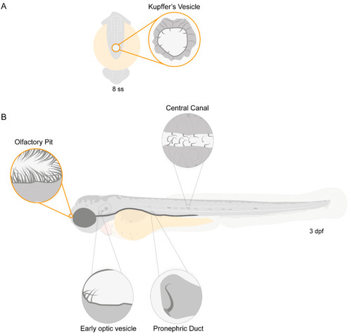
Motile ciliated structures in zebrafish. Schematic representation of (A), a zebrafish embryo at 8 somite stages (ss) highlighting Kupffer’s vesicle (KV); (B) a 3-day post-fertilization (dpf) zebrafish larva indicating structures with motile cilia. Olfactory pit (OP) and KV cilia analysed in the present study are circled in orange. |
Expression Data
Expression Detail
Antibody Labeling
Phenotype Data
Phenotype Detail
Acknowledgments
This image is the copyrighted work of the attributed author or publisher, and
ZFIN has permission only to display this image to its users.
Additional permissions should be obtained from the applicable author or publisher of the image.
Full text @ Int. J. Mol. Sci.