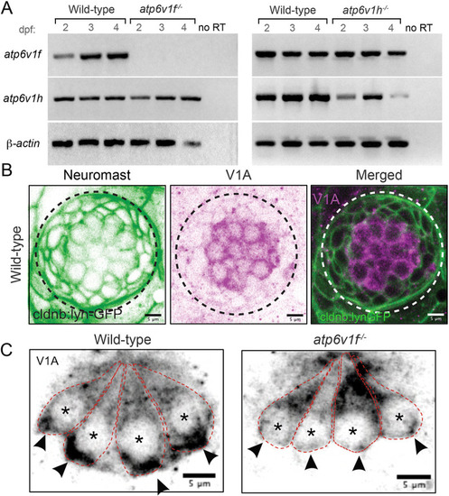
(A) Reverse transcriptase (RT)-PCR analysis of total mRNA from wild-type and V-ATPase mutant embryos at 2, 3 and 4 dpf. atp6v1f mRNA is not detected in atp6v1f−/− embryos, whereas atp6v1h mRNA is detected in the atp6v1h−/− embryos. β-actin mRNA was amplified as a positive control, and reactions without reverse transcriptase (no RT) were negative controls. (B) Antibodies against the V-ATPase V1A subunit (magenta) show enriched staining in centrally localized hair cells in a wild-type neuromast labeled by Tg(cldnb:lynEGFP) expression (green). Dashed line circles indicate the neuromast boundary. (C) Optical sections of hair cells reveal that the V1A subunit localizes throughout wild-type hair cells with an accumulation in the basal region. This basal localization is disrupted in atp6v1f−/− mutant hair cells. Approximate boundaries of individual hair cells are outlined, and asterisks mark hair cell nuclei. Arrowheads indicate basal accumulation of V1A in wild-type hair cells, and lack thereof in atp6v1f−/− hair cells.
|
