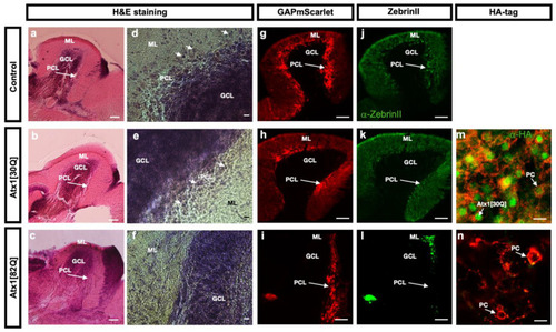
Histological comparison of adult corpus cerebelli of control and hAtx1 expressing zebrafish. Sagittal cryogenic sections through the corpus cerebelli of three months old adult zebrafish from transgenic GAPmScarlet controls (upper row), Atx1[30Q] (middle row) and Atx1[82Q] (lower row) heterozygous carriers, anterior is to the left. (a–c) Hematoxilin and Eosin staining reveals no changes in gross morphology of the corpus cerebelli in overview images, yet at higher magnifications (d–f) large somata (white arrows) characteristic for PCs juxtaposed to the GCL are only visible in control and Atx1[30Q] cerebelli but are hardly found in Atx1[82Q] cerebelli. This absence of PCs is further supported by different patterns of red GAPmScarlet fluorescence, which in controls (g) and Atx1[30Q] (h) samples appears as a continuous layer (white arrow), but can be found in cerebellar tissue of Atx1[82Q] carriers (i) only in posterior regions which is further confirmed by green fluorescent immunohistochemistry using the PC-specific ZebrinII-antibody (j–l). Green fluorescent anti-HA-tag immunohistochemistry confirms the maintenance of human Atx1-protein expression in PCs, yet compared to Atx1[30Q] cerebelli (m) only with sparse labelling in PCs (white arrows) of Atx1[82Q] carriers (n) suggesting a progressive state of degeneration. Scale bars: 100 µm (a–c,g–l), 10 µm (d–f,m,n). Abbr.: GCL: granule cell layer, ML: molecular layer, PCL: Purkinje cell layer.
|
