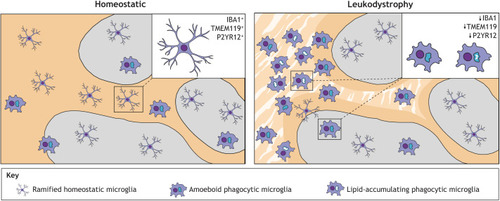
Schematic representation of microglial phenotypes in the grey and white matter of homeostatic and leukodystrophic cortical tissue. In the homeostatic brain, most microglia are evenly distributed in the white and grey matter, and appear ramified, expressing homeostatic markers, such as IBA1, TMEM119 and P2YR12. In the leukodystrophic brain, the white matter is affected by degenerative lesions (striped pattern) and microglia are unevenly distributed, clustering in certain areas, especially within white matter lesions. Phagocytic microglia are abundant within lesions and present an amoeboid shape with large CD68+ intracellular lysosomal vacuoles. Near leukodystrophic lesions, phagocytes display lipid accumulation. Ramified microglia are localized to the grey matter, where their declining density towards the white matter lesions correlates with a gradual loss of homeostatic gene expression and gain of lysosomal CD68 expression. Grey, grey matter; yellow, white matter; stripe pattern, degenerative white matter lesions.
|
