FIGURE SUMMARY
Title

The multicellular interplay of microglia in health and disease: lessons from leukodystrophy

Authors
Berdowski, W.M., Sanderson, L.E., van Ham, T.J.
Source
Full text @ Dis. Model. Mech.

Schematic representation of microglial phenotypes in the grey and white matter of homeostatic and leukodystrophic cortical tissue. In the homeostatic brain, most microglia are evenly distributed in the white and grey matter, and appear ramified, expressing homeostatic markers, such as IBA1, TMEM119 and P2YR12. In the leukodystrophic brain, the white matter is affected by degenerative lesions (striped pattern) and microglia are unevenly distributed, clustering in certain areas, especially within white matter lesions. Phagocytic microglia are abundant within lesions and present an amoeboid shape with large CD68+ intracellular lysosomal vacuoles. Near leukodystrophic lesions, phagocytes display lipid accumulation. Ramified microglia are localized to the grey matter, where their declining density towards the white matter lesions correlates with a gradual loss of homeostatic gene expression and gain of lysosomal CD68 expression. Grey, grey matter; yellow, white matter; stripe pattern, degenerative white matter lesions.

Leukodystrophy-associated proteins in microglia. Several microglial proteins, either at the cell surface or residing within organelles, are involved in leukodystrophy-related cellular processes. CSF1R is a dimeric transmembrane receptor that is a key regulator of microglia and macrophage biology, including proliferation, migration and survival, and binds ligands CSF-1 and IL-34. TREM2 is a single-pass transmembrane receptor involved in lipid metabolism and phagocytic clearance by macrophages and microglia. It binds anionic ligands, including phospholipids and bacterial components, and forms a signaling complex with DAP12, which is a dimeric transmembrane adapter required for the cell-surface expression of TREM2, forming a signaling complex. LRRC33 is a leucine-rich repeat-containing protein that anchors latent TGF-β1 at the cell surface. It is required for activation of the TGF-β1 pathway in macrophages and microglia. The USP18 isopeptidase binds to the intracellular domain of IFNAR2, thereby negatively regulating the IFN-I pathway. ALDP, a peroxisomal ABC half-transporter encoded by ABCD1, transports VLCFAs. ASA, encoded by ARSA, is a lysosomal enzyme that digests sulfatides, glycosphingolipids that are highly enriched in myelin. GALC, encoded by GALC, is a lysosomal enzyme catabolizing galactosylceramide and psychosine, both major glycosphingolipids in myelin. ASA and GALC are not known to be expressed at high levels in macrophages and microglia, but there are strong indications that their functions are required by microglia in the context of myelin.
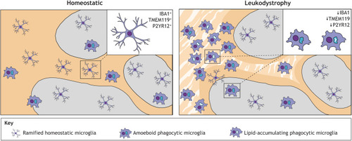
Loss of microglial functions could lead to white matter degeneration by affecting multiple intercellular connections. Routes whereby microglia, directly or indirectly, affect the white matter (WM) have been supported by data from experimental studies as described here; however, their relevance to disease remains to be fully explored. Astrocytes compensate for a lack of microglial phagocytosis by becoming more phagocytic, although they are less efficient than microglia and this response may result in the neglect of critical astrocytic functions. Together with the increased astrocytic reactivity observed in leukodystrophies, this can result in disturbed lipid and metabolic supply to oligodendrocytes (OL) and an unsupportive ECM environment for OPCs. Additionally, altered interactions between the BBB and astrocytic end-feet can perturb metabolic supply to brain cells. Aberrant microglia can lead to insufficient trophic support for OPCs and oligodendrocytes, and a diminished oligodendrocyte lineage. Aberrant microglia may also exhibit perturbed clearance and pruning capacity and contribute to impaired remyelination. Both aberrant microglia and affected astrocytes can cause neuronal stress due to neurotoxicity, ineffective phagocytosis and/or dysregulation of neuroactivity. Axonal pathology and abnormal neuronal activation can affect myelination and, in turn, the degeneration of myelin results in a loss of metabolic support for axons. In sum, white matter degeneration in leukodystrophies is likely preceded by distinct effects of aberrant microglia, possibly forming a ‘perfect storm’ of parallel effects particularly detrimental for the myelinated white matter tracts. Solid arrows indicate established interaction/consequence. Dashed arrows indicate hypothesized interaction/consequence in leukodystrophic brain.

Acknowledgments
This image is the copyrighted work of the attributed author or publisher, and ZFIN has permission only to display this image to its users. Additional permissions should be obtained from the applicable author or publisher of the image. Full text @ Dis. Model. Mech.