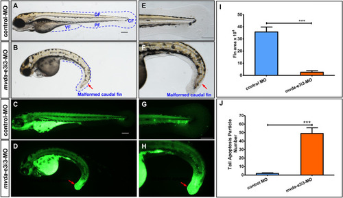
mvda deficiency induces fin-reduction phenotypes and tail-specific apoptosis. A normal fins, control-MO; B fin-reduction phenotypes after mvda-e3i3-MO injected; E–F caudal fin is shown at higher magnification. Dashed lines indicate the morphology of fins. Control-MO-injected embryos and embryos injected with mvda-e3i3-MO were stained with acridine orange at 4-dpf. Apoptotic cells are visible as bright green spots, and less bright homogenous green or black staining is unspecific background staining. C, G Control-MO-injected zebrafish exhibited few or no apoptotic cells in whole organism. D, H In contrast, significantly increased staining was observed throughout the tail in zebrafish injected with mvda-e3i3-MO (red arrows). I Quantification of area at fin shows a 13.9-fold decrease in mvda morphants. Error bars, s.e.m.; ***P < 0.0001(n = 10; ANOVA). J Quantification of apoptosis particle number at tail shows a 25.8-fold increase in mvda morphants. Error bars, s.e.m.; ***P < 0.0001(n = 10; Student’s t test); A–H: lateral view, anterior, left. CF caudal fin, DF dorsal fin, PF pelvic fin, VF ventral fin, dpf days post fertilization. Scale bars = 100 µm
|
